Astrocytic Hamartoma Mimicking Cotton Wool Spot
Purpose. The aim of this paper is to describe the key features of astrocytic hamartoma that distinguish it from cotton wool spots, the most common differential diagnosis among white retinal findings.
Material and Methods. A case report of a 35-year-old female patient with a history of type 2 diabetes mellitus and primary hypertension is presented as a framework for the differential diagnosis of astrocytic hamartoma. Ultra-widefield retinal imaging and optical coherence tomography (OCT) of findings from an additional patient are presented for comparison.
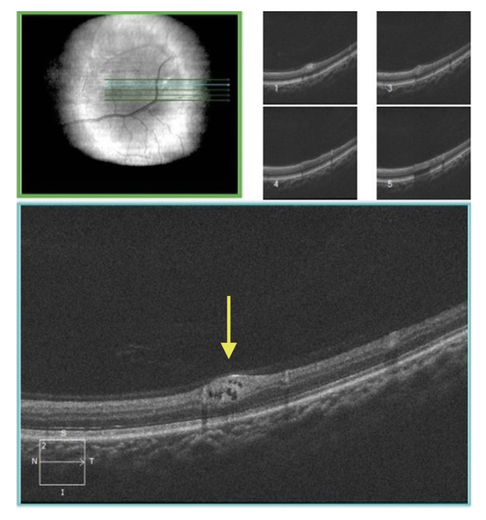
Results. The diagnosis of astrocytic hamartoma was established after comprehensive dilated fundus examination and high-definition OCT. A five-line raster OCT scan over the lesion demonstrated a hyperreflective, slightly elevated mass with rounded hyporeflective cystic spaces spanning from the retinal nerve fiber layer to the inner nuclear layer, which excluded the previously assigned diagnosis of cotton wool spot. Review of fundus photographs from prior examinations documented slow lesion growth, providing additional support for the final diagnosis.
Conclusion. Astrocytic hamartoma should be included in the differential diagnosis whenever a white retinal lesion is encountered, particularly in diabetic and hypertensive patients in whom cotton wool spots are a feature of progressive retinopathy. Key distinguishing criteria are lesion location relative to the optic nerve head, stability or slow growth over time, and characteristic OCT features, including hyperreflectivity and cystic spaces.
Introduction
Astrocytic hamartomas are benign retinal tumors arising from glial cells. They may closely resemble cotton wool spots (CWSs), the most clinically important differential diagnosis among white retinal findings, making accurate diagnosis challenging during routine clinical examination.1,2 Astrocytic hamartomas typically present as white or yellowish-white, slightly elevated lesions located proximal to the optic nerve head, although disc involvement can also occur.2,3 They are classified as either calcified and nodular or non-calcified and flat, and they grow in an exophytic pattern, projecting outward from the inner retinal surface rather than extending inward into the underlying tissue.1
Lesions tend to be slow-growing, and regression has been reported.2 Astrocytic hamartomas may occur as isolated, idiopathic findings in otherwise healthy individuals, but they are also strongly associated with tuberous sclerosis complex (TSC), an autosomal dominant neurocutaneous disorder characterized by hamartomatous growths in multiple organ systems.4,5,6 In patients with TSC, multiple retinal hamartomas are commonly observed bilaterally.6
Cotton wool spots represent focal areas of axoplasmic flow stasis within the retinal nerve fiber layer (RNFL) secondary to ischemia.1 They are common findings in diabetic retinopathy, hypertensive retinopathy, and other systemic vascular conditions.
Optical coherence tomography (OCT) has become an indispensable tool in differentiating these white retinal lesions.2 This report is not intended to describe a novel disease entity. Rather, its primary value lies in providing an instructive diagnostic lesson that illustrates the differential diagnosis of white retinal lesions through multimodal imaging. This case report presents a patient with concurrent diabetes mellitus and hypertension in whom an astrocytic hamartoma was initially misdiagnosed as a cotton wool spot, and discusses the clinical and imaging features essential for accurate differentiation.
Case description
A 35-year-old female presented to our clinic for a routine comprehensive eye examination. Her most recent prior examination, performed one year earlier by an outside provider, had documented a diagnosis of myopia (both eyes) and moderate non-proliferative diabetic retinopathy (left eye [OS]). Her systemic medical history was significant for type 2 diabetes mellitus, primary hypertension, and hyperlipidemia. Directed dermatologic and neurologic review of systems was negative for cutaneous manifestations associated with tuberous sclerosis complex (e.g., hypomelanotic macules, facial angiofibromas, shagreen patch) and for a history of seizures or developmental delay. At the time of presentation, her most recent glycated hemoglobin (HbA1c) was 8.0 % and in-office blood pressure measured 130/95 mmHg.
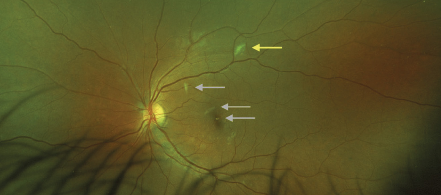
Entrance testing, including entering and best-corrected visual acuity and cover test, was unremarkable. Anterior segment findings on slit-lamp biomicroscopy were within normal limits. Dilated fundus examination revealed an opaque, grossly flat, white retinal lesion located at the bifurcation of the superior temporal arcade in the left eye (Figure 1). The lesion had been documented at the prior examination and was recorded as a cotton wool spot. All other posterior segment findings were unremarkable and consistent with the previous examination records.
A high-definition five-line raster OCT scan was performed to characterize the lesion further (Figure 2). OCT imaging demonstrated a primarily hyperreflective, slightly elevated lesion with rounded hyporeflective cystic spaces involving the retinal nerve fiber layer through to the inner nuclear layer. These findings were inconsistent with a cotton wool spot, which would be expected to appear as a focal area of hyperreflectivity confined to the RNFL with no cystic spaces.1,2 The patient was tentatively diagnosed with a non-calcified astrocytic hamartoma and was instructed to return in three months for monitoring.
At the three-month follow-up visit, the lesion demonstrated no clinically appreciable growth compared with the prior examination; OCT findings were consistent with the baseline scan. The findings were communicated to the patient’s primary care provider. Given the benign, slow-growing nature of the lesion, the patient was placed on annual monitoring.
Fundus images from prior examinations, retrieved as far back as 19 months before the index visit (September 2020), were obtained and reviewed. Comparison of the lesion across these time points confirmed a gradual, slow increase in lesion size over the documented 19-month period, further supporting the diagnosis of astrocytic hamartoma over a transient ischemic lesion such as a cotton wool spot, which would be expected to resolve within weeks to months. Representative serial fundus photographs documenting this gradual increase in lesion size over this 19-month observation period are presented in Figure 3.
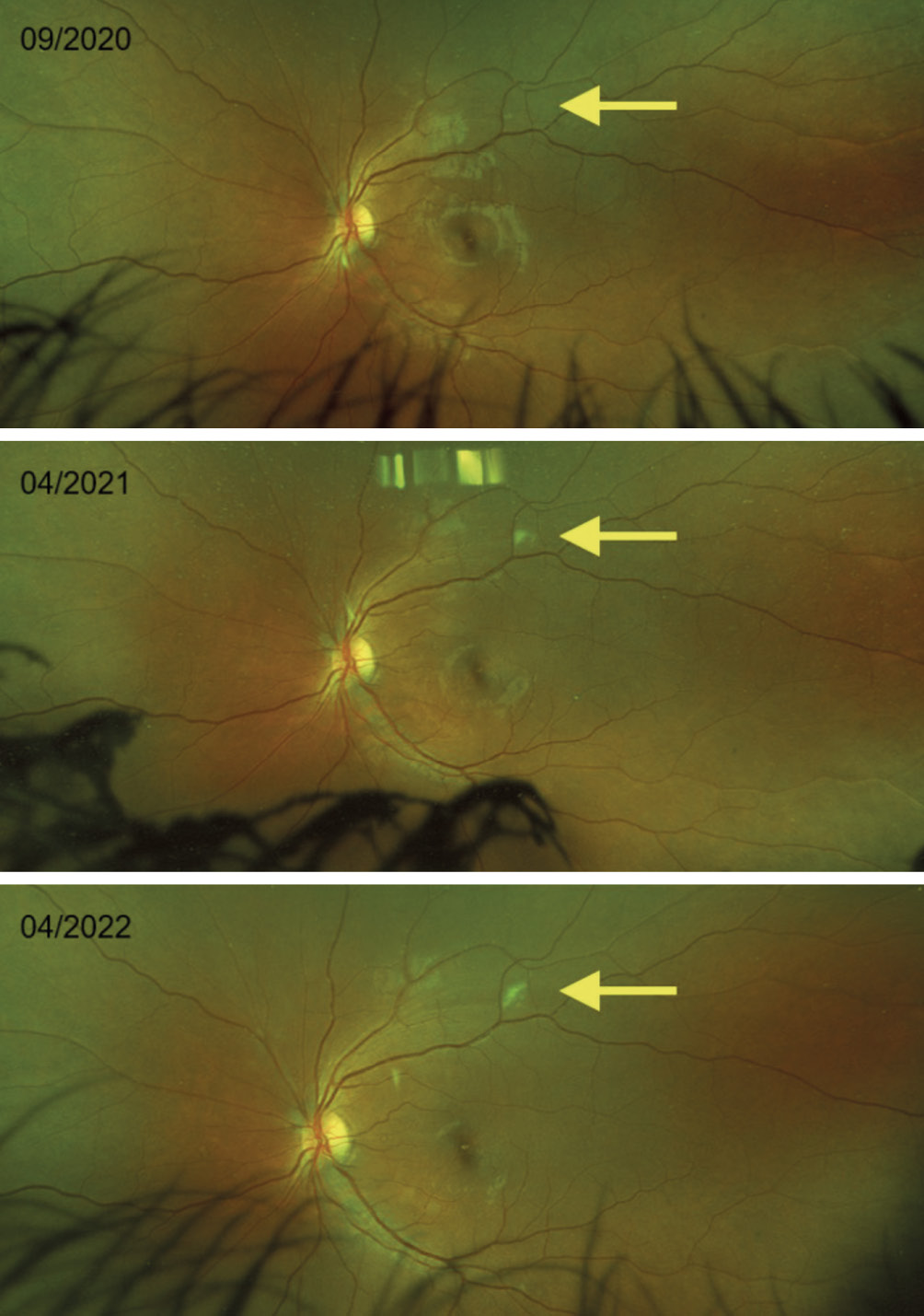
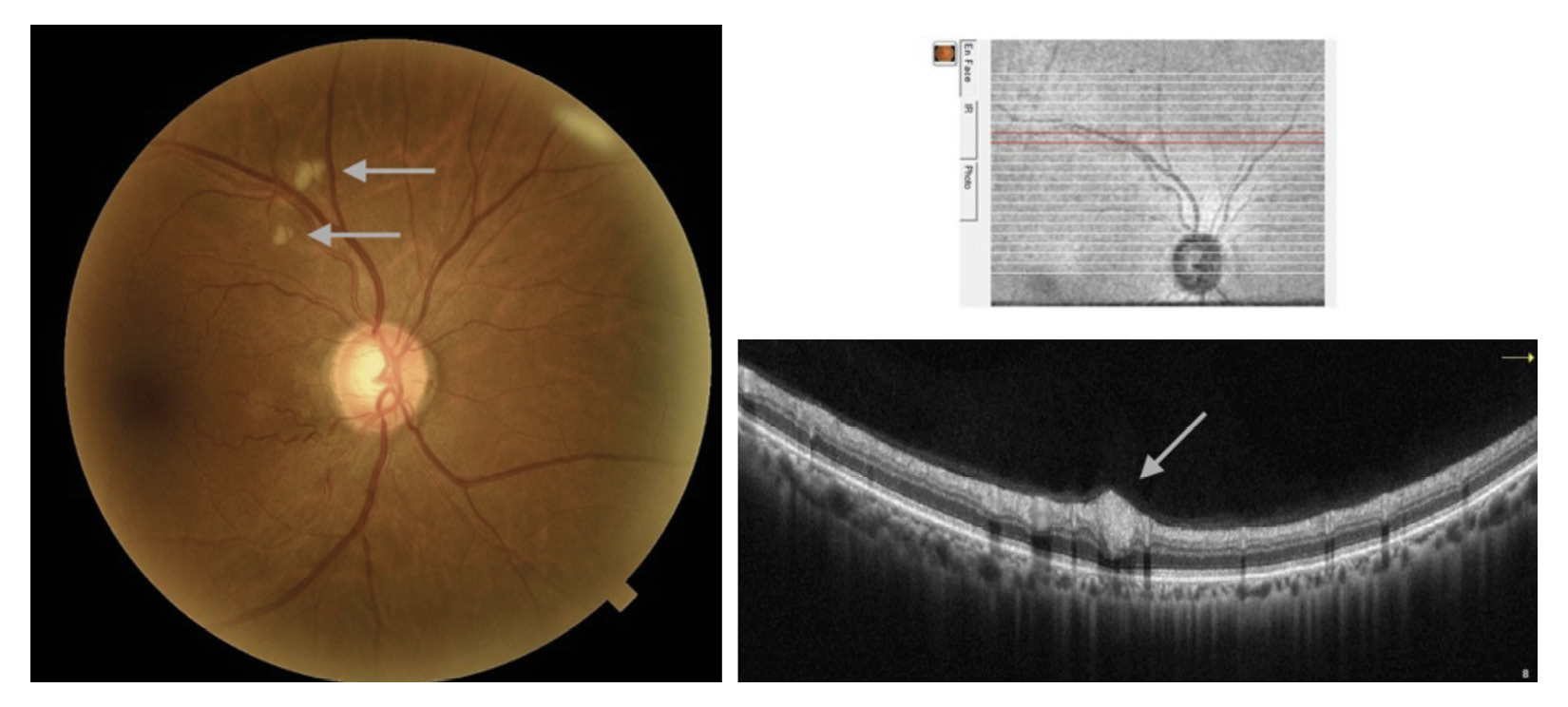
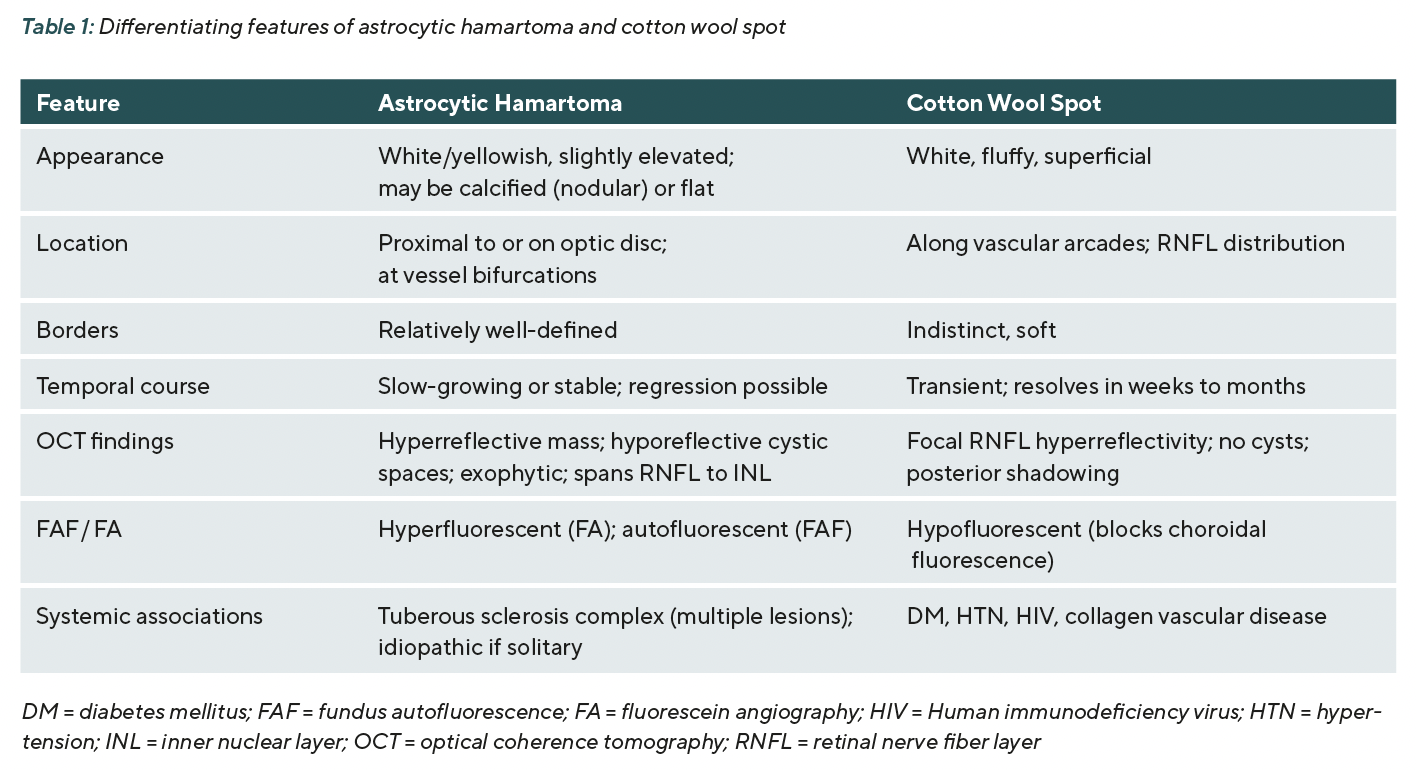
Table 1 summarizes the clinical and OCT features that distinguish astrocytic hamartoma from cotton wool spot. A representative widefield fundus image and OCT scan illustrating cotton wool spot (Figure 4) from a different clinic patient is presented for comparison.
Discussion
Astrocytic hamartomas are rare but clinically significant retinal lesions that may be misidentified as more common conditions in patients with predisposing systemic disease.2,5 The case presented here illustrates precisely this diagnostic pitfall: a white retinal lesion in a diabetic, hypertensive patient was attributed to the more prevalent condition, cotton wool spot, for at least one year before OCT prompted reconsideration.
The key to the correct diagnosis in this case was the OCT appearance. Whereas cotton wool spots produce focal, superficial RNFL hyperreflectivity without structural deformation of deeper retinal layers, astrocytic hamartomas demonstrate an exophytic, hyperreflective mass with characteristic hyporeflective cystic spaces that can extend from the RNFL to the inner nuclear layer.2,4 This OCT signature has been described in prior series and is considered highly characteristic.2,4,6 Additional recent reports have further refined the OCT-based characterization of astrocytic hamartoma, including a morphologic classification system correlating tomographic subtype with systemic tuberous sclerosis complex manifestations,9 as well as more recent work demonstrating the value of OCT in detecting subtle lesions not apparent on fundus photography.10 The cystic spaces visible on OCT likely represent areas of mucinous degeneration or fluid accumulation within the hamartoma.1
Location is another important differentiating feature. Astrocytic hamartomas arise from retinal astrocytes and are therefore found along the course of retinal nerve fibers, most commonly adjacent to the optic disc or at vessel bifurcations within the posterior pole.2,6 Cotton wool spots, by contrast, are distributed along the vascular arcades and correspond to areas of capillary non-perfusion. While location alone is insufficient to distinguish these entities, the combination of proximity to the disc, persistence on serial imaging, and characteristic OCT appearance is highly supportive of hamartoma.
The temporal course of the lesion proved critical in this case. Cotton wool spots are transient ischemic infarcts of the RNFL that typically resolve within 4–12 weeks as axoplasmic debris is resorbed.1 Persistence or growth of a white retinal lesion beyond this timeframe should raise suspicion for an alternative diagnosis. Here, retrospective review of fundus photographs confirmed that the lesion had been present and slowly enlarging for at least 19 months, a timeline entirely inconsistent with a cotton wool spot.
Fluorescein angiography (FA) may provide additional diagnostic information when the diagnosis remains uncertain after OCT. Astrocytic hamartomas are typically hyperfluorescent on FA due to intrinsic staining and leakage from tumor vasculature.1,2 Cotton wool spots, in contrast, block background choroidal fluorescence and therefore appear hypofluorescent. Fundus autofluorescence (FAF) may reveal autofluorescence of the hamartoma, which has been attributed to lipofuscin-like material within the lesion.6 These additional imaging modalities were not performed in the index case, but should be considered when the OCT findings are equivocal.
When an astrocytic hamartoma is identified, whether solitary or multiple, the possibility of tuberous sclerosis complex must be considered. TSC is an autosomal dominant multisystem disorder caused by mutations in the TSC1 or TSC2 genes and is associated with hamartomas in the brain, kidneys, skin, lungs, and retina.5,6 Retinal hamartomas occur in approximately 40–50 % of TSC patients and may be the presenting manifestation of the syndrome.5 Solitary retinal hamartomas in otherwise healthy patients, so-called idiopathic astrocytic hamartomas, have also been described.6,7 In the present case, the patient had a single lesion with no personal or family history suggestive of TSC; therefore, she did not meet diagnostic criteria for TSC and the hamartoma was considered idiopathic. Although the vast majority of astrocytic hamartomas, including the lesion described here, follow a benign and slow-growing course, rare cases have been reported to grow aggressively. These aggressive cases most often arise in the setting of tuberous sclerosis complex, with associated release of vascular endothelial growth factor (VEGF) leading to choroidal or retinal neovascularization and secondary neovascular glaucoma.8 This rare but vision-threatening potential underscores the importance of continued periodic monitoring even in lesions that initially appear clinically stable. Nonetheless, appropriate systemic screening history and neurological review of systems should be obtained for all patients in whom a retinal astrocytic hamartoma is newly identified.
When located on the optic disc, astrocytic hamartomas must be further distinguished from optic nerve head (ONH) drusen, which can also appear as refractile, calcified nodules at the disc surface.2 Calcified astrocytic hamartomas and ONH drusen share a similar white, glistening appearance and may both exhibit autofluorescence; however, B-scan ultrasonography and OCT can help differentiate them, as drusen typically occupy a subsurface position within the disc substance, whereas calcified hamartomas project from the disc surface.
Management of astrocytic hamartomas is generally conservative. Because lesions are benign and slow-growing, and because no effective treatment is available or necessary for most cases, annual dilated fundus examination with photographic documentation is appropriate.2,6 Patients should be advised of the nature of the lesion and the importance of follow-up. Systemic workup for TSC should be initiated if clinical features suggest the possibility.
Conclusions
Astrocytic hamartoma is a benign retinal lesion that can closely resemble cotton wool spots and other white retinal findings, particularly in patients with diabetes mellitus or hypertension. In such patients, a white retinal lesion that fails to resolve within the expected timeframe for a cotton wool spot, or that demonstrates characteristic OCT features such as hyperreflectivity, hyporeflective cystic spaces, and exophytic morphology, should prompt consideration of astrocytic hamartoma as the diagnosis. High-definition OCT is the most useful single modality for differentiating these entities in clinical practice. Reviewing longitudinal fundus photography is invaluable when available. Once diagnosed, hamartomas should be monitored annually and, if multiple, should prompt evaluation for tuberous sclerosis complex.
Conflict of interest
The authors declare that there is no conflict of interests regarding the methods and devices mentioned in the article.
COE Multiple Choice Questionnaire
The publication "Astrocytic Hamartoma Mimicking Cotton Wool Spot" has been approved as a COE continuing education article by the German Quality Association for Optometric Services (GOL). The deadline to answer the questions is 1st November 2027. Only one answer per question is correct. Successful completion requires answering four of the six questions.
You can take the continuing education exam while logged in.
Users who are not yet logged in can register for ocl-online free of charge here.