The influence of age on the intraocular pressure of the human eye
Purpose. The aim of this study was to investigate whether age has a significant influence on intraocular pressure and central corneal thickness. Due to conflicting results from previous studies, this study aims to provide new impetus for clarifying these correlations.
Material and Methods. The retrospective, quantitative, non-interventional cross-sectional study included 190 subjects from an optometry practice in the Seychelles (2025) and 237 subjects from previosly unpublished survey data collected by Petermann and Wildgrube in Germany (2010). In the Seychelles, intraocular pressure and central corneal thickness were measured using a non-contact tonometer. In Germany, intraocular pressure was also measured with a non-contact tonometer, whereas central corneal thickness was assessed using optical coherence tomography (OCT). Statistical analysis was performed separately for both locations.
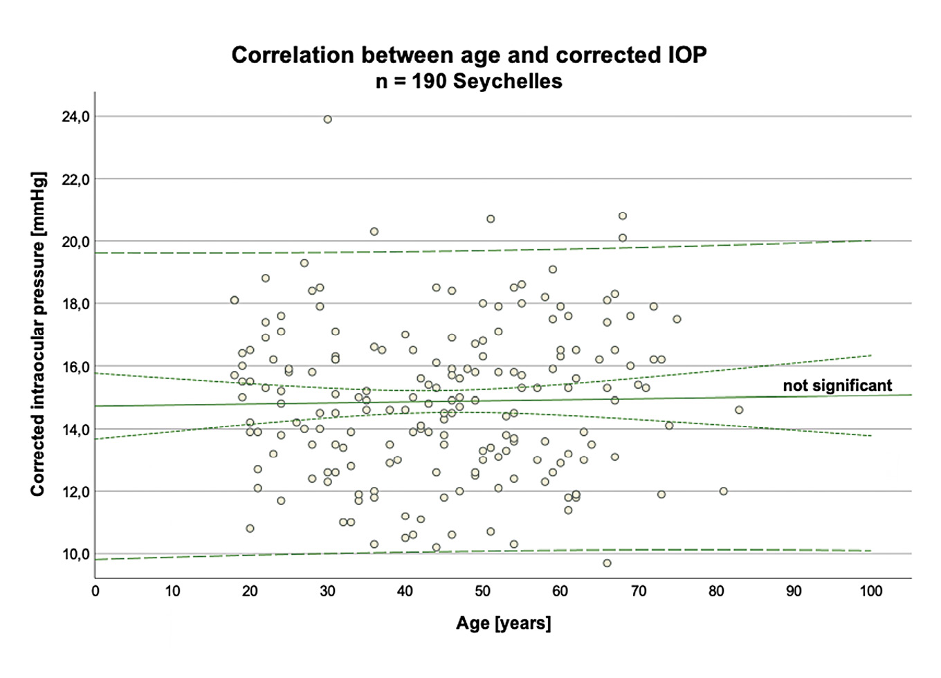
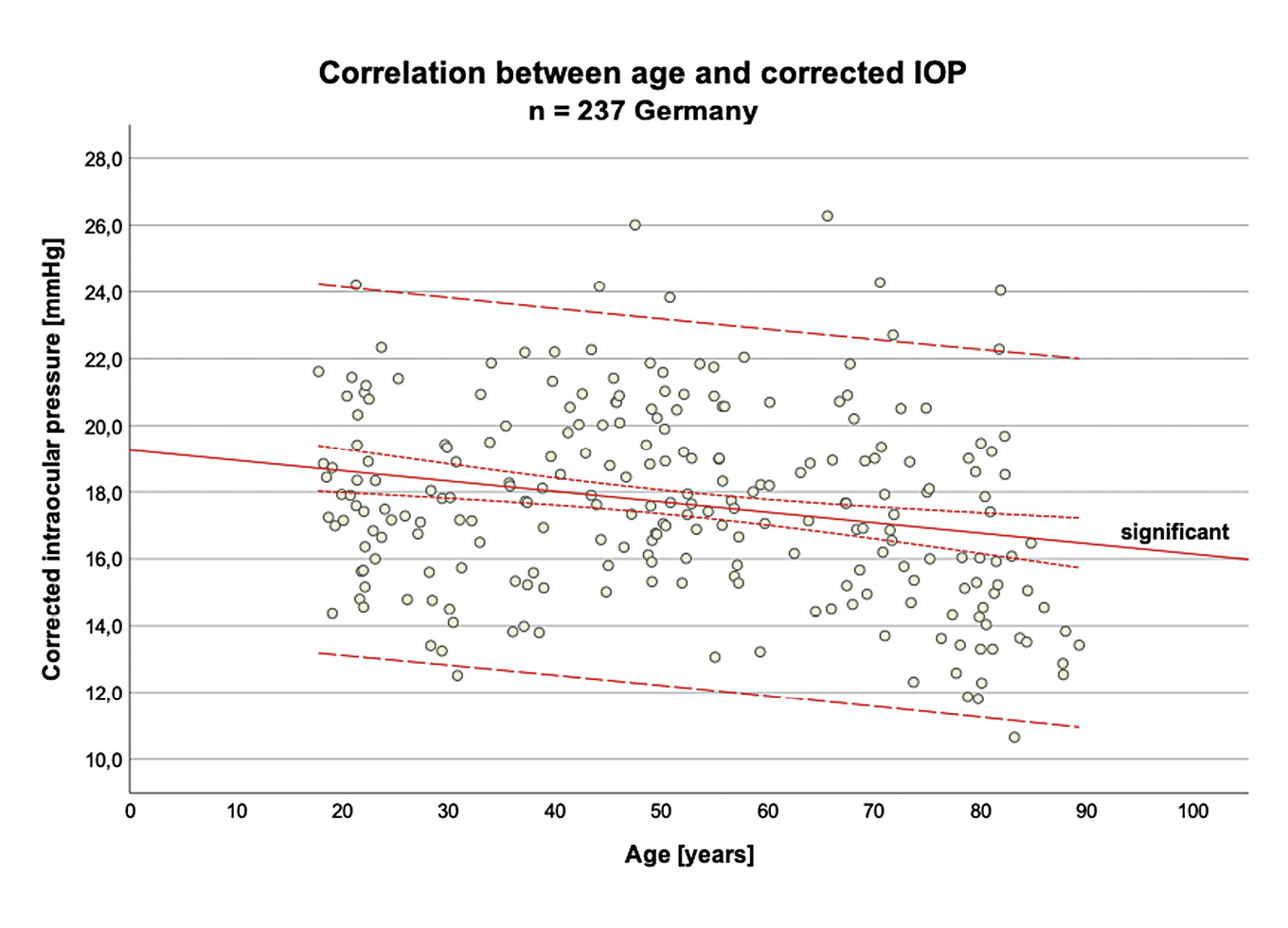
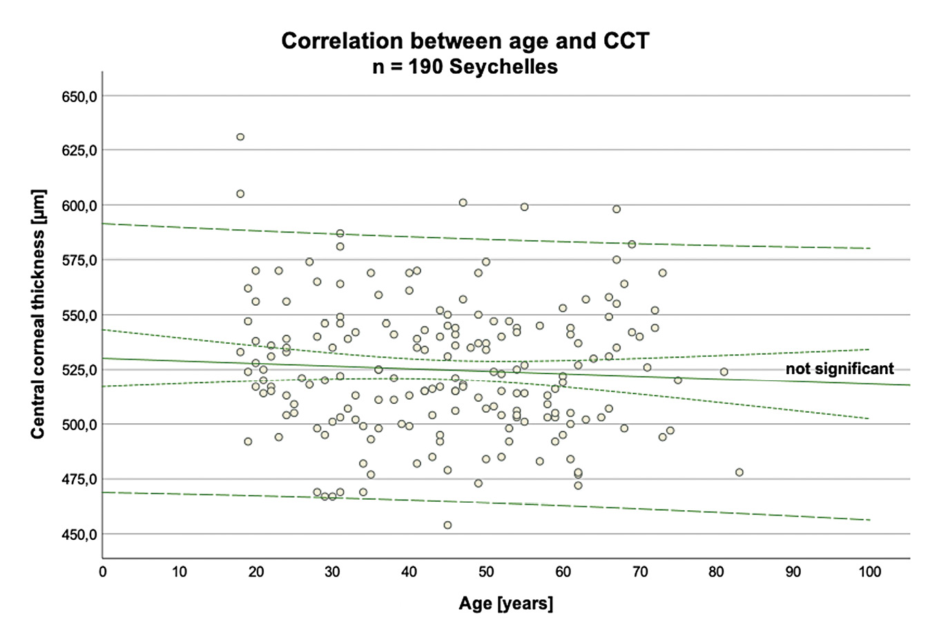
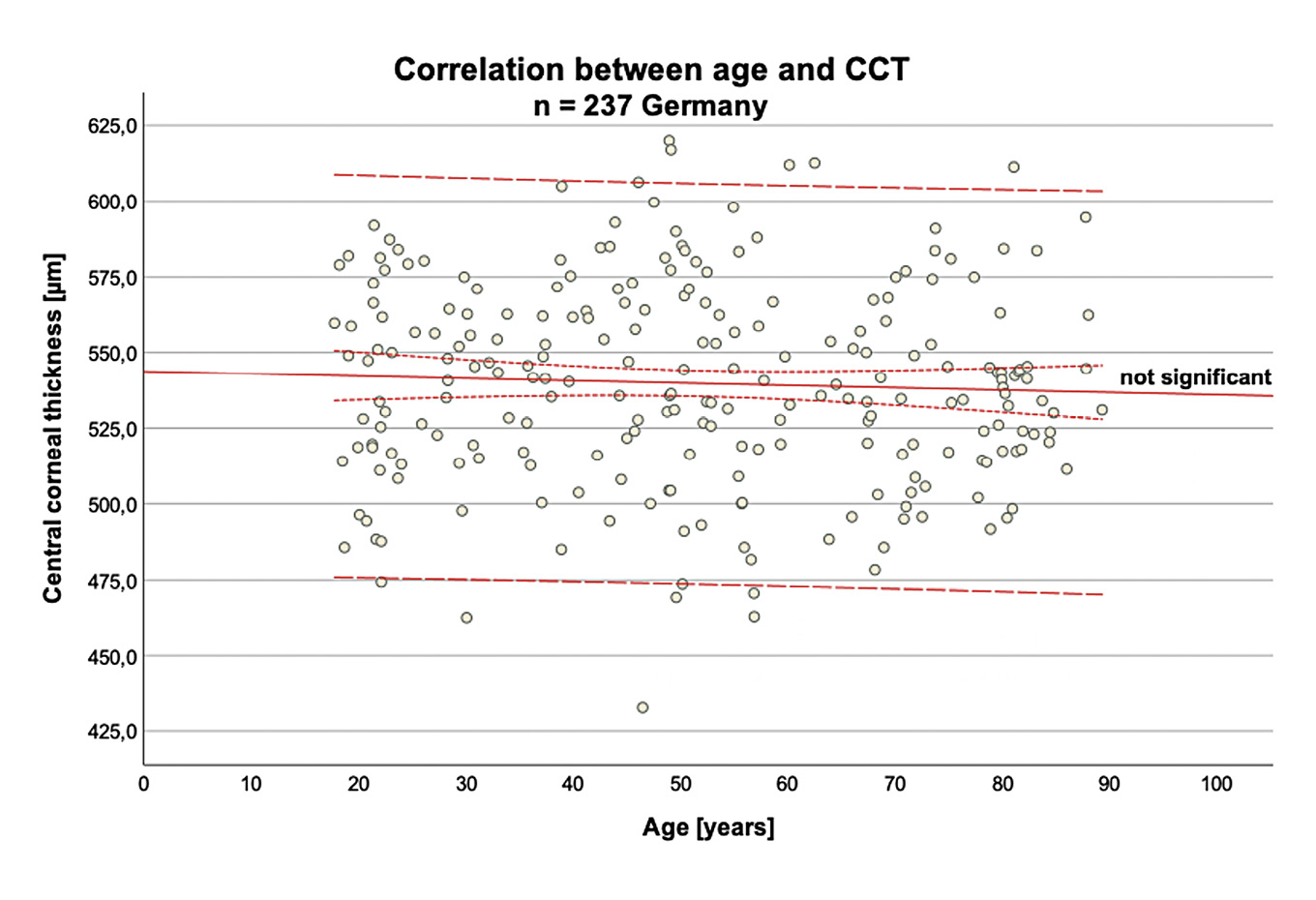
Results. A significant negative correlation between age and intraocular pressure was found only in the German data (p < 0.001; r = −0.226), while no significant correlation was found in the Seychelles (p = 0.709; r = 0.027). There was no significant correlation between age and central corneal thickness at either location (Seychelles p = 0.506; r = −0.049; Germany p = 0.287; r = −0.069).
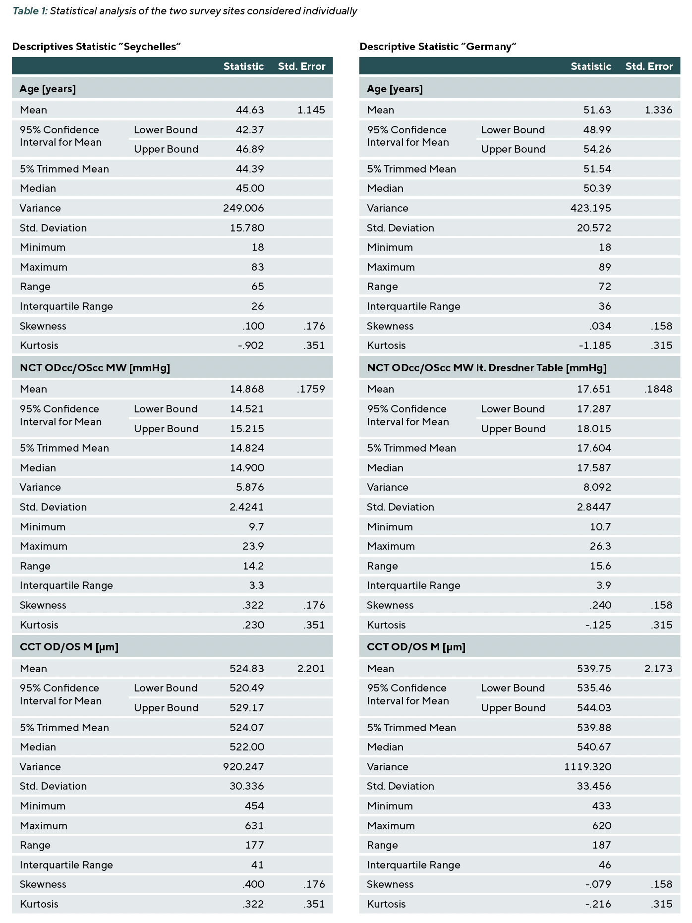
In the Seychelles, the average age was 44.63 years (SD ± 15.78), the mean intraocular pressure was 14.87 mmHg (SD ± 2.42), and the central corneal thickness was 524.83 µm (SD ± 30.34). In Germany, the average age was 51.63 years (SD ± 20.57), the mean intraocular pressure was 17.65 mmHg (SD ± 2.84), and the central corneal thickness was 539.75 µm (SD ± 33.46).
Conclusion. The correlation between age and intraocular pressure may vary depending on the population. While a significant negative correlation was found in Germany, this was not found in the Seychelles. These results imply that the age-related increase in primary open angle glaucoma cannot be explained by a general increase in intraocular pressure with age. No significant correlation between age and central corneal thickness was found at either location.
Introduction
As we age, structural and functional changes occur in the eye that can affect intraocular pressure (IOP). In particular, degenerative processes in the trabecular meshwork and the uveoscleral drainage pathway lead to a reduced rate of aqueous humour outflow, which can contribute to an increase in intraocular pressure. These age-related changes are clinically significant, as elevated intraocular pressure is considered a major risk factor for the development of glaucoma. This condition involves irreversible damage to the optic nerve and, if left untreated, is one of the most common causes of blindness worldwide.
Intraocular pressure is not, however, a constant value, but is subject to a variety of influencing factors, including genetic factors, diurnal fluctuations and systemic parameters. Age, in particular, is frequently discussed as a possible influencing factor, although there is no clear scientific consensus.1,2 Whilst some studies document an increase in IOP with advancing age,3,4,5,6 others show a decrease 7,8,9,10 or no significant association.11,12,13,14
Central corneal thickness (CCT) is also important for the interpretation of intraocular pressure. Age-related changes have been described, although there is no consistent body of evidence. Several studies have reported an age-related decrease in CCT, although the strength of this association varied between different populations.15,16,17 Ethnic differences have also been described, including a lower CCT in individuals of African descent compared with Caucasian controlgroups.18,19,20
In addition to biological factors, the measurement accuracy and reliability of the instruments used also influence the assessment of statistical correlations. Device-specific differences between instruments have been described in non-contact tonometry.21 Limited measurement accuracy and/or reproducibility can lead to fluctuations in the measured intraocular pressure values and thereby influence the assessment of statistical correlations.22,23
Against this background, the present study investigated the relationship between age and intraocular pressure. Additionally, the relationship between age and central corneal thickness was examined. The aim was to analyse age-related changes, identify possible trends and determine their significance for the clinical assessment of intraocular pressure and CCT.
Materials and methods
Study design
The study design was a retrospective, quantitative, non-interventional cross-sectional study covering the geographical locations of the Seychelles and Germany. For the Seychelles location, we used data provided by a specialist optometric practice (‘VisionCare Seychelles’) originating from 2025; for the German location, data from a previously unpublished Bachelor’s thesis [Petermann and Wildgrube] were used.24 The study was conducted in accordance with the ethical principles of the Declaration of Helsinki, taking into account the European General Data Protection Regulation and the guidelines of the Berlin University of Applied Sciences (BHT).To ensure the comparability of the data, standardised measurement procedures were employed. Calibrated and validated instruments were used, multiple measurements were taken and averaged, and an identical mathematical correction of intraocular pressure was applied.
Inclusion and exclusion criteria
The study included participants aged 18 years or older who were able to maintain sufficient fixation. Participants with systemic conditions such as diabetes or high blood pressure were considered suitable and included, as it can be assumed that whilst these conditions may influence intraocular pressure, they are not uncommon in the general population and exclusion would have introduced unwanted bias to the study.
Any participants with known glaucoma and individuals taking IOP-lowering medication were excluded, due to their impact on intraocular pressure. Individuals were also excluded where measuring IOP did not appear feasible due to corneal surgical or pathological changes and their impact on the biomechanics of the cornea.
Participants
After applying the aforementioned inclusion and exclusion criteria, n = 190 participants with an average age of 44.63 years (SD ± 15.78) were included in the study for the Seychelles site. It was not possible to elicit the ethnic groups in detail, as the population of the Seychelles consists of ethnically diverse and mixed ethnic groups.
At the German site, n = 237 participants with an average age of 51.63 years (SD ± 20.57) and predominantly of Caucasian ethnicity were included in the study.
Equipment
To measure intraocular pressure and corneal thickness at the Seychelles site, the non-invasive non-contact tonometer (NCT) known as the Tonoref III (Nidek Co Ltd, Gamagori, Japan) was used. This device measures the uncorrected IOP three times per eye and calculates an average value from these readings. In addition, the central corneal thickness (CCT) is measured and presented as an average value, which is also derived from three measurements per eye. This allows the corrected IOP value to be calculated. This is done via an automated calculation within the device using the Dresden correction table, which is based on the mean value of the uncorrected IOP and takes the measured CCT into account.
In Germany, a non-contact tonometer designated AT 555 (Reichert Inc, Depew, NY, USA) was also used for the non-invasive measurement of intraocular pressure. This device also measures the uncorrected IOP value three times per eye and calculates an average value from these.
The Visante OCT (Carl Zeiss Meditec AG, Jena, Germany) was used to measure central corneal thickness.24 As the device measures CCT only once, three measurements were taken consecutively and an average value was calculated from these.
To ensure the data were comparable, the uncorrected IOP values were manually corrected using the Dresden correction table. The conversion was performed using the following formula:
IOP(corrected [mmHg])
= IOP(uncorrected [mmHg]) + (550 [µm] − CCT [µm]) / (25 [µm])
Statistical methods
The statistical analyses were performed using the IBM SPSS Statistics software version 29 (IBM, Chicago, IL, USA) on the ‘macOS’ operating system (Apple Inc, Cupertino, CA, USA).
Descriptive statistics and exploratory data analysis were carried out on all collected data to facilitate analysis and verify data quality. The Shapiro-Wilk test was used to assess normality of distribution. Histograms and QQ plots were also used for visual inspection.
A correlation analysis was performed to investigate the relationship between age and corrected intraocular pressure, as well as between age and central corneal thickness. Where at least one of the two variables was not normally distributed, the non-parametric Spearman’s test was selected. The correlation test was conducted as a two-tailed test.
A regression analysis was performed only if a correlation existed.
To better illustrate the relationships, scatter plots were created showing regression lines, the 95 % confidence interval and the 95 % prediction interval.
To ensure better comparability, a significance level of α = 0.05 was set for all statistical tests, with a p-value < 0.05 being considered statistically significant.
Results
The results of the statistical analysis for the two survey sites are examined individually below. All data used are based on mean values (Table 1).
The correlation analysis between age and the adjusted IOP
For the geographical location Seychelles, the correlation analysis revealed no association (r = 0.027; p = 0.709) (Figure 1).
Consequently, the alternative hypothesis (H₁) – that age influences intraocular pressure in the human eye – could not be confirmed.
When considering data from Germany, a significant correlation was found (r = −0.226; p < 0.001). Accordingly, the corrected IOP tends to decrease slightly with increasing age (Figure 2).
To describe the relationship between the two variables in greater detail, a regression analysis was also performed. A statistically significant correlation was found (r² = 0.051; p < 0.001). The standard deviation of the residuals was ± 20.09 years. The regression coefficient (B = −1.63; SE = 0.46) indicates that, in the present sample, older age was associated with lower (corrected) intraocular pressure. Accordingly, younger subjects had, on average, higher values of corrected intraocular pressure. However, it should be noted that there is considerable variation in the data, and the predictive power associated with this should therefore be considered limited.
The correlation analysis between age and CCT
For the Seychelles location, this test revealed no correlation (r = −0.049; p = 0.506) (Figure 3). A similar result was observed when analysing the data from Germany with no correlation being observed (r = −0.069; p = 0.287) (Figure 4).
Discussion
In this study, data from two geographical locations, the Seychelles and Germany, were analysed separately. The aim was to investigate possible associations between age, intraocular pressure and central corneal thickness on a population-specific basis.
For the Seychelles sample, there was no evidence of an age-related association with either intraocular pressure or central corneal thickness.
Based on the German sample, a weak but statistically significant association was noted between age and intraocular pressure. Surprisingly, higher age was associated with lower intraocular pressure. However, the model accounted only for a small proportion of the variance. Similarly, there was no association between age and corneal thickness in the German population.
These findings are partly consistent with previous studies while confirming the mixed evidence regarding an age-related effect on IOP. Whilst large-scale studies such as the ‘Barbados Eye Study’ 3, the ‘Beaver Dam Eye Study’ 4 and other studies 5,6 report a slight age-related increase in IOP, long-term studies in Asia in particular 7,8 as well as cross-sectional studies 10, such as the ‘Japan Ningen Dock Study’ 9, show an age-related decrease in IOP. Interestingly, non-contact tonometry was used throughout these Asian studies, whereas Goldmann applanation tonometry was predominantly used in the Western studies. The choice of measurement method could therefore have a significant influence on the observed results. Equally, the ethnic composition of the populations studied suggests differing IOP values in relation to age and suggests population-specific differences in ocular, systemic or genetic factors as possible explanations.
Some studies,12,13,14 including the ‘Blue Mountains Eye Study’ 11 from Australia, found no association between age and IOP. The results of these studies highlight the importance of a nuanced consideration of the findings, taking into account potential confounding factors such as systemic disease, family history, and ethnic background. In particular, the ‘Blue Mountains Eye Study’ showed that an initially observed age-dependent increase in IOP resulted in a negative correlation after systemic diseases were excluded. After further adjustment for ocular diseases, the results no longer showed any age-related association. This observation is somewhat in contrast with the findings of the present study, in which IOP-relevant ocular diseases were excluded (but included systemic diseases). However, a statistically significant but weak negative trend was observed only in the German sample and there was no consistent age-dependent effect on IOP.
The ‘Baltimore Eye Survey’ 25 and the ‘South African Eye Study’ 18 demonstrated that ethnicity can influence IOP. It was found that Africans had, on average, a higher IOP than the comparison group, which included Caucasians. These findings was also inconsistent with our study, as the sample from the Seychelles yielded lower mean IOP values (M 14.87 mmHg) than the German sample (M ≈ 17.65 mmHg).
When interpreting these findings, the specific ethnic characteristics of the Seychelles’ population must be taken into account. A clear differentiation of the individual ethnic groups was not possible in the present study, as the indigenous population is highly mixed. The current population structure reflects historical settlement by French colonists, as well as by African, British, Indian, and Chinese groups. Despite its geographical affiliation with Africa, the population of the Seychelles cannot therefore be regarded as ethnically homogeneous. This may limit comparability with study populations of exclusively African origin.
Similarly, central corneal thickness is influenced by both age and ethnicity, as various studies 16,17,18,19,20 have shown. For instance, the ‘Tehran Eye Study’ 15 from Iran reported, in a large population-based study, an age-related decrease in CCT, with older subjects tending to have a thinner cornea. This could not be confirmed in our study, as no significant correlation could be demonstrated in either the sample from the Seychelles or the German population. These findings could be interpreted as indications that the cornea does not undergo clinically relevant changes with age, but the discrepancy to previous studies has not been resolved. Comparing CCT in ethnic groups, the ‘South African Eye Study’ 18 documented significant ethnic differences, with individuals of African descent having, on average, a thinner central cornea than Caucasian individuals. These findings are also reflected in the results of the present study, in that the sample from the Seychelles showed a tendency towards a lower CCT (M = 524.83 µm) compared to the German study population (M = 539.75 µm), although the difference was small. This finding can also be explained by the mixed ethnic structure of the Seychelles’ population.
The diversity of the existing research literature can be explained not only by methodological and population-specific factors, but also by differences in study design. Whereas longitudinal studies typically yield more robust insights into age-related trends, cross-sectional studies, such as the present study, provide only a snapshot in time and may be affected by structural biases. It also became apparent that certain age groups visited the optometry practice more frequently, resulting in an uneven age distribution within the sample. To address this issue in future research, targeted recruitment aimed at achieving a more balanced age distribution is recommended.
Another potential methodological limitation concerns the measuring devices used. Non-contact tonometers from different manufacturers were used at both geographical locations. Although these devices employ broadly standardised procedures, device-specific differences may result in slight variations in measurements. However, we consider any potential instrument error to be of relatively minor importance for the interpretation of the correlation analysis, as such errors are systematic and would affect all measured values within a sample equally. In future studies it may be more accurate to measure all intraocular pressure (IOP) values using the same non-contact tonometer.
It should also be noted that the medical and ocular history were self-reported by participants, i.e. no clinical records were reviewed for verification purposes. This leaves a residual risk of data inaccuracies. To mitigate, clearly defined inclusion and exclusion criteria were used while considering central corneal thickness as a potential confounding variable, which facilitated a reliable and clinically relevant data set.
The findings of this study provide guidance for clinical practice. The results support current clinical practice that clinical decisions, such as the diagnosis of glaucoma, should not be solely based on an any individual clinical variable such as expected age-related trend in IOP or CCT. Rather, an individual interpretation of the measurements and risk assessment is required for each patient, taking into account a multitude of factors including age, central corneal thickness, pre-existing systemic conditions, history of ocular and systemic diseases, and ethnic origin. This individualised approach is consistent with established clinical practice, as even larger studies have not consistently demonstrated a universally reliable correlation between age and the parameters examined. The present findings are therefore in line with the existing body of evidence.
For future studies, it would be advisable to ensure a balanced age distribution. Studies could also be extended to include additional populations and ethnic groups in order to better capture potential differences and to further assess the generalisability of the findings. It may also be beneficial to include the measurement of blood pressure as a variable to reduce the impact of any potential misreporting by participants and associated bias.
Additional factors such as corneal biomechanics, refractive status, axial length and individual lifestyle factors could be considered to achieve a more nuanced understanding of IOP development across the lifespan. It may be valuable to examine ocular conditions in a more differentiated manner in order to better assess their potential effects.
In summary, the present findings, together with parts of the international literature, suggest that age-related correlations between intraocular pressure and central corneal thickness cannot be described consistently and may, in some instances, only be weak. Against this background, alongside population-specific influences, it should be considered that a stable and universally applicable correlation may not be present.
Conclusion
In this study, potential associations between age, intraocular pressure and central corneal thickness were examined separately in two different populations. In the Seychelles sample, no significant age-related association were observed for either IOP or CCT. In contrast, the German sample showed a significant albeit weak association between age and IOP. However, the low proportion of explained variance suggests limited explanatory power. No significant association between age and CCT was oberserved.
Overall, the results are consistent with the heterogenous findings reported in the literature and highlight the influence of methodological, population-specific and ethnic factors. The findings suggest that age-related changes in IOP and CCT cannot be assumed uniformly. In clinical practice, measurements should therefore be interpreted on an individual basis, considering relevant influencing factors. The results contribute to a more nuanced understanding of age-related correlations across populations.
Conflict of interest
The authors have no conflict of interest regarding the methods and equipment mentioned in the article.