A Novel ZNF408 Variant in Recessive Retinitis Pigmentosa
Purpose. The purpose of this paper is to document a mutation of the ZNF408 gene not noted in existing literature in a patient with phenotypic presentation of retinitis pigmentosa (RP).
Material and Methods. A 27-year-old Asian male of Bangladesh descent presented with a concern of night vision difficulty. He had no significant medical or ocular history. Fundus examination revealed bone spicules in the peripheral retina of both eyes. Imaging was conducted utilizing Optos ultra-widefield imaging with fundus autofluorescence [(FAF), Optos Daytona, USA], Zeiss Optical Coherence Tomography [(OCT), Zeiss, Germany] of the retina and optic nerves, and 30-2 Humphrey visual fields (HVF) in both eyes. A full-field electroretinogram (ffERG) was performed to examine rod and cone function. A buccal swab was collected and the Invitae Inherited Retinal Disorders Panel was ordered.
Results. Fundus photography documented bone-spicule
pigmentation in the peripheral retinas of both eyes. OCT of the optic nerve showed bilateral thickening of the retinal nerve fiber layer. OCT of the macula revealed parafoveal thinning of the photoreceptors in both eyes. FAF imaging revealed bilateral parafoveal hyper-autofloursence. HVF 30-2 testing showed superior arcuate defects in both eyes. ffERG results indicated reduced photopic and scotopic responses. Genetic testing revealed a homozygous mutation of uncertain significance on the ZNF408 gene.
Conclusion. Genetic testing is a valuable tool in the diagnosis of RP and other inherited retinal disorders. However, understanding of the genetics involved with hereditary retinal disorders remains incomplete. Reporting cases of phenotypic presentations of disease with variants of uncertain significance increases collective understanding of the genetics of RP.
Introduction
Retinitis pigmentosa (RP) is an inherited retinal disease that is characterized by the degeneration of the rod and cone photoreceptors. RP can be inherited as autosomal dominant, X-linked, or most commonly as autosomal recessive.1 The initial symptoms of RP include being unable to see well in dim light settings or situations that require fast adjustments from light to dim environments, usually presenting at around 20 years of age.1 Eventual decreased peripheral vision often becomes more apparent over time but is typically not initially noted.1
Clinical findings include the “classic triad” seen on funduscopy evaluation: bone spicule pigmentation, vascular narrowing, and waxy pallor of the optic nerve.1 These signs may not initially present early in the disease and the degree to which these signs present do not necessarily correlate with the severity of the disease.1 Other common secondary findings include posterior subcapsular cataracts and cystoid macular edema.1
Over 90 genes have been associated with syndromic and non-syndromic RP, but a mutation does not guarantee presence of the disease. Conversely, the absence of mutations in genes known to be associated with RP does not rule out the diagnosis, as our understanding of RP genetics continues to evolve.2
Material and Methods
Ultra wide field fundus photography with FAF was performed using Optos Daytona (Optos, United States). Scans from the Zeiss Optical Coherence Tomography was used to assess the retinal nerve fiber layer and other retinal layers (Zeiss, Germany). Peripheral visual fields were assessed using the 30-2 Humphrey Visual Field (Zeiss, Germany). Rod and cone responses were obtained with the ffERG (Diagnosys, United States) performed under photopic (cone response) and scotopic (rod response) settings. Genetic testing was conducted via buccal swab with the Invitae Inherited Retinal Disorders panel (Labcorp, United States).
Results
The patient’s best corrected visual acuity was measured as 20/20 in the right eye and 20/20 in the left eye with his habitual contact lenses. Pupils were equal, round, and reactive with no presence of an afferent pupillary defect in the right or left eye. Confrontational visual fields were full to finger counting in the right and left eye. Extraocular muscle motility was full in both eyes. Intraocular eye pressures were measured at 18 mmHg in each eye.
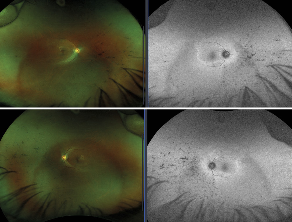
Anterior segment evaluation showed no remarkable findings. A dilated fundus examination revealed perfused optic nerves with distinct margins and a flat macula in each eye. The retinal blood vessels appeared to be attenuated in each eye. The peripheral retina had pigmentation in the shape of bone spicules 360 degrees, most concentrated inferiorly in both eyes. Otherwise, there were no holes, breaks, or tears in the peripheral retina in both eyes.
Retinal photos confirmed the bone spicule pigmentation in the peripheral retina, more noticeable inferiorly. FAF imaging showed bilateral paramacular hyper-autofluorescence (Figure 1).
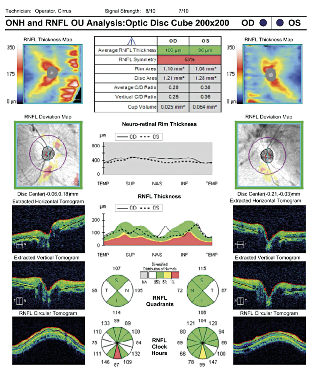
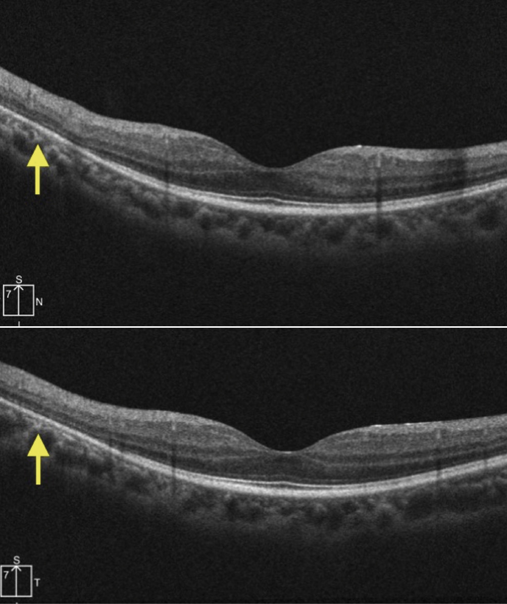
OCT of the optic nerve showed a thickened retinal nerve fiber layer compared to the normative database nasal and temporal OD and temporal OS. This is indicated by the white regions in the RNFL Quadrants section of the OCT (Figure 2). OCT of the macula revealed bilateral parafoveal thinning of the photoreceptor integrity line in both eyes with intact subfoveal retinal architecture (Figure 3).
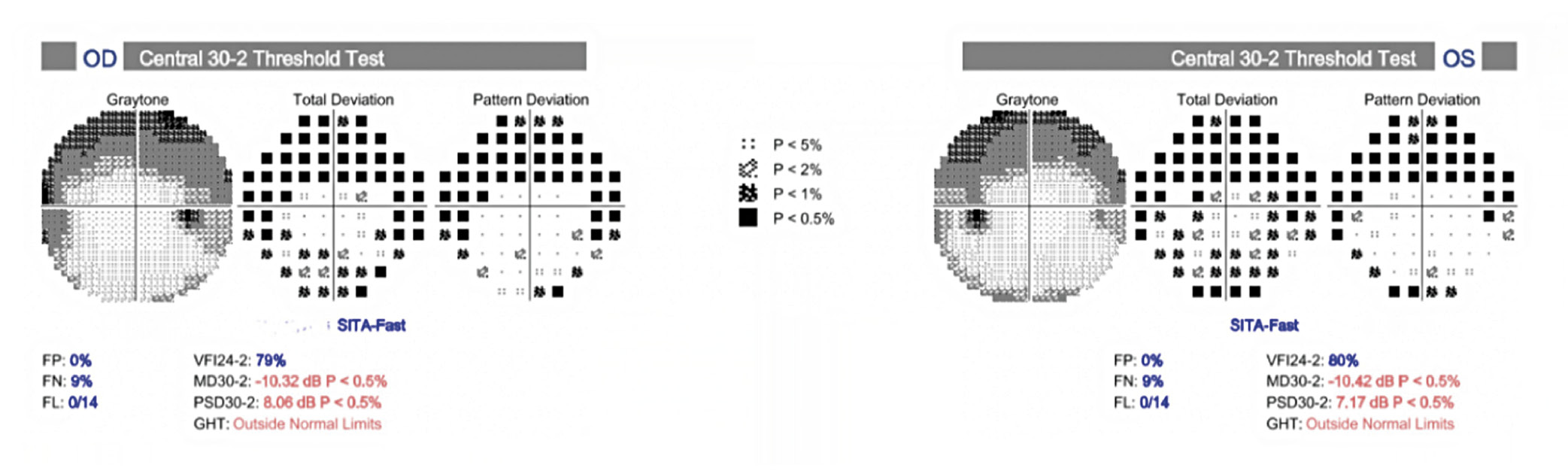
The 30-2 Humphrey visual fiend showed a superior arcuate defect in both eyes, which corresponded to the hyper-autofluorescence in the inferior paramacular region seen in the retinal FAF. The intensity of the hyper-autofluorescence does not necessarily correlate directly to the severity of the visual field loss. The hyper-autofluorescence in the paramacular region is more pronounced in the right eye, however the visual field shows similarly dense defects, if not a slightly more advanced defect in the left eye (Figure 4). As with visual acuity, structure may not always mirror function and it should be considered that FAF imaging is an objective test, while the visual field is subjective and thus open to increased
variability.
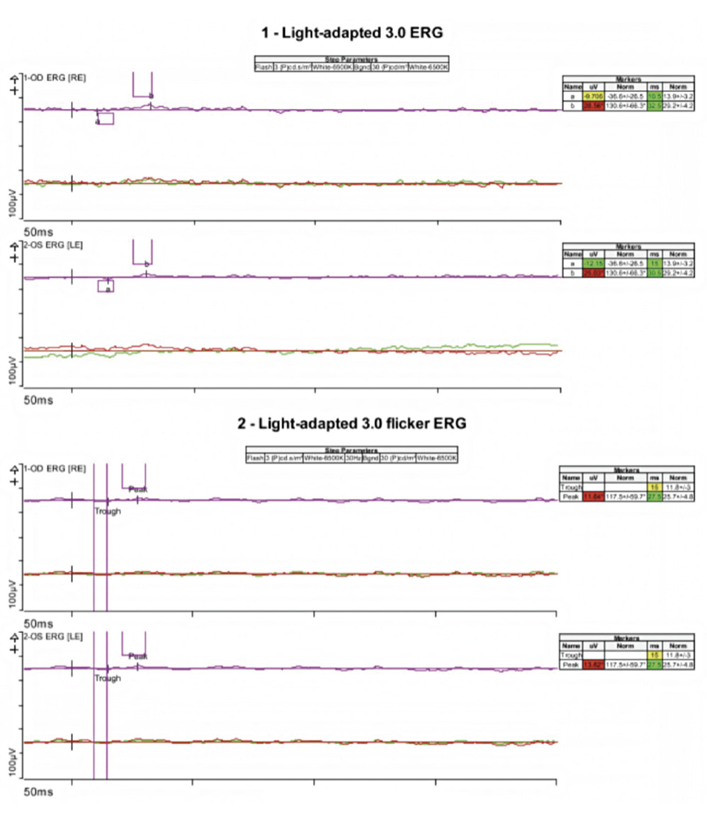
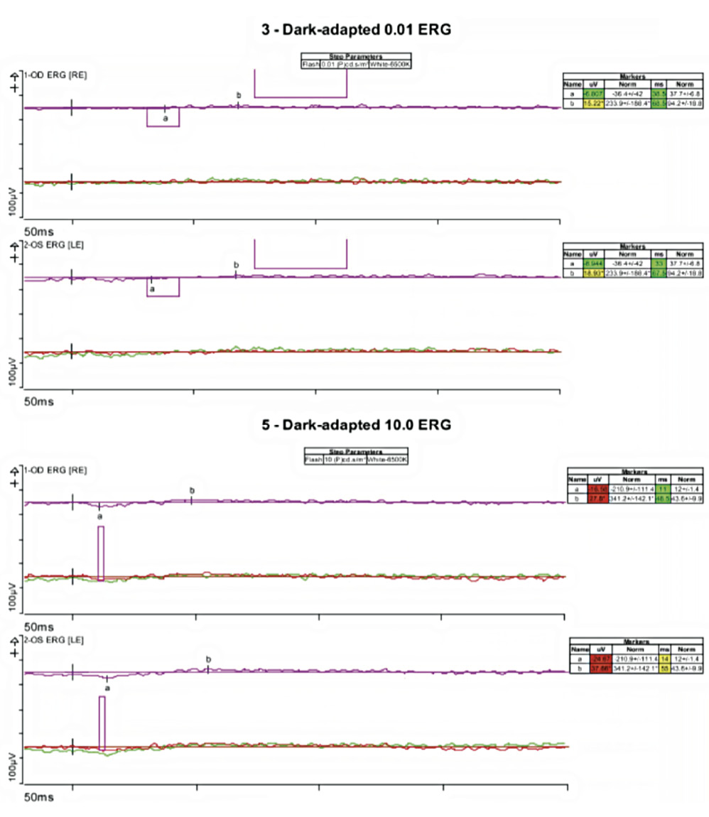
The dark adapted 0.01 ERG (rod response) and the dark adapted 10.0 ERG rod-cone maximal response were essentially extinguished (Figure 5). The light adapted 3.0 ERG demonstrated a significantly reduced response in the right eye and left eye. The light-adapted 3.0 flicker ERG was essentially extinguished for both eyes (Figure 6). These responses were typical of advanced RP.
Genetic testing revealed a homozygous mutation in gene ZNF408, Exon 5. The mutation replaced glycine with aspartic acid at codon 419. The report indicated that this variant has not been reported in the literature in individuals with ZNF408-related conditions.
Discussion
The patient had clinical signs consistent with a diagnosis of advanced retinitis pigmentosa. He demonstrated bone spicule pigmentation in the peripheral retina and his arterioles were attenuated in appearance. The appearance of a hyper-autoflourescent parafoveal ring (Robson-Holder ring) on FAF imaging is indicative of stress of the parafoveal retinal pigment epithelium (RPE). Malfunctions in the RPE results in increased lipofuscin accumulation and is responsible for the hyper-autofluorescent appearance. A Robson-Holder ring is associated with a spectrum of inherited retinal diseases and photoreceptor dysfunction.3 The Robson Holder ring corresponded to attenuation of the photoreceptor integrity line seen inferiorly on OCT imaging and a superior arcuate visual field defect, indicating more photoreceptor loss inferiorly in both eyes.
Thickening of the retinal nerve fiber layer in patients with retinitis pigmentosa has been observed particularly in the temporal quadrants.4 One characteristic often observed in retinitis pigmentosa is a “waxy pallor” of the optic nerve without atrophy. One proposed hypothesis for this observation is that damage to the outer retinal layers in RP triggers astrocytic gliosis. However, literature examining the RNFL variability in patients with RP is inconsistent and further study is needed.
The ffERG confirmed the diagnosis given the extinguished rod responses (dark adapted) relative to cone responses (light adapted), which were also extremely reduced. Typically, in retinitis pigmentosa, rod function is affected earlier than cone function, but cone function is also reduced or extinguished as the disease progresses.5 The patient’s results suggest damage to both the rod and cone photoreceptors. It should be noted that, as in the present patient, ffERG results may be reduced or extinguished but the patient may still exhibit normal visual acuity due to the survival of some remaining cones.
The ZNF408 gene is associated with autosomal recessive retinitis pigmentosa.6 However, the mutation identified was reported as a variant of uncertain significance, meaning the association between this mutation and disease has not been firmly established in the literature. The patient’s mutation had glycine, a neutral and non-polar amino acid, switched with the negatively-charged and acidic aspartic acid.7 The Poly-Phen 2 predictive algorithm 8 for genetic mutations predicts that this substitution is likely to be disruptive to the functioning of the gene due to the contrasting amino acid properties.
Retinitis pigmentosa has a high prevalence in certain South Asian communities, where consanguineous marriages are common. Consanguinity is often associated with the recessive disease and has been linked to the higher occurrence of RP in Southern India.9 Thirty-two out of the 60 known genes thought to be involved with non-syndromic RP were identified in South Asia. At a tertiary care referral center in Southern India, every sixth patient with a positive family history of RP was found to have a history of consanguineous marriage. However, the available literature remains limited.10
Conclusion
Genetic testing is a valuable tool in the diagnosis of RP and other inherited retinal diseases. However, our understanding of the genetics involved with these hereditary retinal disorders remains incomplete. Performing testing on individuals, even if they have the typical presentation of the disease, is essential in contributing to the database and increasing our knowledge of the role the genetics play.
Conflict of Interest
The authors declare that there are no conflicts of interests regarding the methods and devices mentioned in the article.