Red Light in Myopia: Short-Term Effects of Direct LED Red Light on Subfoveal Choroidal Thickness and Axial Length
Purpose. The aim of this study was to investigate short-term effects of direct LED red light on subfoveal choroidal thickness (SfChT) and axial length (AL).
Material and Methods. In this experimental, randomized controlled crossover study 37 healthy adults aged 24 ± 2 years were recruited. Participants were randomized into two groups. Each participant was exposed to two different light stimuli in two separate experimental sessions for 20 minutes: a) LED red light (623 ± 17 nm, 0.0040 mW/mm², 1000 lx), and b) LED white light (6500 K, 0.0035 mW/mm², 1000 lx). The sessions were conducted at two different appointments at similar times of day with an interval of one to two weeks. SfChT and AL of the right eye were measured before and immediately after light exposure.
Results. Using paired t-test there were no significant differences between baseline and after LED red or white light exposure in SfChT (mean change after red light: +1 ± 10 μm, p = 0.758; after white light: 0 ± 8 μm, p = 0.878) and AL (mean change after red light: 0.00 ± 0.01 mm, p = 0.542; after white light: 0.00 ± 0.01 mm, p = 0.279). Furthermore, there were no significant differences between red and white light exposure (mean difference in SfChT: 0 ± 10 μm, p = 0.854; AL: 0.00 ± 0.02 mm, p = 0.249).
Conclusion. The investigated LED stimuli showed no short-term effect on SfChT and AL. Further research is needed to determine whether, and under what conditions, exposure to red LED light can have a clinical effect on axial length and choroidal thickness and thus may have a therapeutic effect in the context of myopia control.
Introduction
In recent decades, the prevalence of myopia has increased worldwide.1 In Germany, approximately 35 % to 40 % 2 and in parts of Asia, over 90 % of adults are individuals with myopia. It is predicted that in the coming years and decades, even more people will become myopic.1 There has been an increase in the number of children with myopia not least due to the COVID-19 pandemic.3 Several risk factors have been investigated in relation to the development of myopia, ranging from genetics, to light exposure, binocular vision and the biomechanics of the eye.4–6 Myopia in children is often associated with excessive axial elongation of the globe.7 This causes the retina, choroid, and sclera to stretch and thin, which can lead to various complications.8,9 Studies have suggested that choroidal thickness may be related to axial length change with thinning of the choroid being associated with increased axial length, whereas thickening of the choroid was associated with decreased axial length.10,11 Therefore, the choroid could be an indicator for the development and progression of myopia and a target for therapeutic methods.12 Recent studies have revealed that alterations in choroidal thickness and choroidal blood flow play important roles in the progression of myopia. Reduced choroidal blood flow could affect scleral cellular matrix remodeling, which leads to axial elongation and further myopia progression.13
In this context, the International Myopia Institute Report on the choroid highlights the question of whether short-term changes of the choroid could be a biomarker for future ocular growth.14 Moreover, several studies have shown that the time spent outdoors has an inhibitory effect on the development of myopia.15–18 Therefore, it is reasonable to assume that light is a decisive factor in the regulation of eye growth. Some animal experiments, for example, with tree shrews and rhesus monkeys, showed that long-wavelength red light in the visible spectrum or long-pass filters for red light induced hyperopia, reduced axial length, and increased choroidal thickness.19–21 Consistent with these results, several randomized controlled trials have been published since 2021, investigating a potential new therapy with (Repeated) Low-Level Red Light (RLRL/LLRL) to inhibit axial growth in progressive myopia.22,23 RLRL involves irradiating the eyes twice daily for three minutes using a laser that generates long-wavelength red light in the visible spectrum with a power of up to 2 ± 0.5 mW. This treatment resulted in a significant reduction in axial length and an increase in choroidal thickness.24–26 However, there are concerns regarding the safety of this therapy. Ostrin and Schill reported that the recommended safety limits may have been exceeded.27 In addition, a case report described a possible association between red light laser therapy and retinal damage in a 12-year-old patient.28
Light emitting diodes (LEDs) may have effects similar to lasers, particularly in the long-wavelength visible spectrum, since they can emit wavelengths comparable to RLRL devices.29 However, available RLRL devices differ from LED red light, for example, in terms of irradiance and light source size. Lou and Ostrin 30 and Thakur et al.31 reported that one hour of ambient LED red light (630 or 623 nm) significantly thinned the choroid in human subjects. This was observed with a comparatively lower intensity than in conventional RLRL therapy and animal experiments, and contrasts with the results of those studies. Therefore, the question remains as to whether the high intensity of the light, the wavelength alone, the interaction of both, or other causes affect choroidal thickness and axial length, and thus, the regulation of eye growth in humans. Based on the current study situation, the aim of this study was to investigate the short-term effect of LED red light with higher intensity through direct illumination on choroidal thickness and axial length.
Material and Methods
Study Design and Participants
This experimental, randomized controlled crossover study was conducted between October 2023 and November 2023 at the Ernst Abbe University of Applied Sciences Jena (EAH Jena). The study was conducted in accordance with the principles of the Declaration of Helsinki and approved by the Ethics Committee of the Medical Faculty of Friedrich Schiller University Jena in October 2023. Written informed consent was obtained from all participants prior to enrollment.
Participants in the study were students and employees of the EAH Jena. Inclusion and exclusion criteria were selected based on parameters from similar studies.30,31 Inclusion criteria were age between 18 and 40 years, a spherical equivalent (SE) of −6.00 D to +6.00 D to rule out high ametropia, and a best-corrected visual acuity of 0.10 logMAR or less of the right eye. Exclusion criteria were eye diseases, systemic diseases or medications that could affect vision and current or previous therapies regarding myopia management.
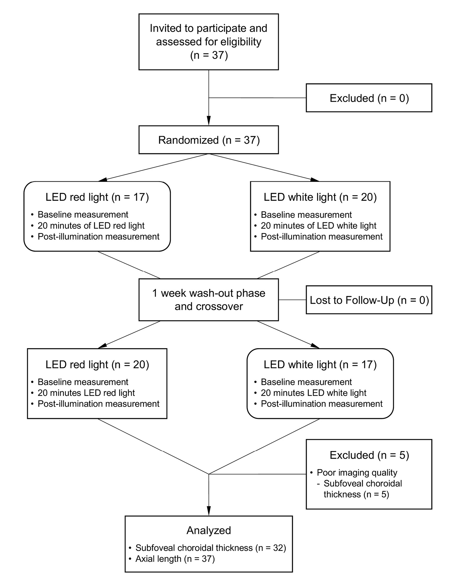
Each participant was invited to two examination appointments on different days between 8 a.m. and 4 p.m. The two examinations were carried out at similar times of day to avoid the influence of diurnal fluctuations as these can affect axial length and choroidal thickness.32–34 Attention was also paid to short-term effects on choroidal thickness, such as exercise, caffeine, and alcohol.14 On both study days, an initial assessment of the outcome measures was conducted, followed by 20 minutes of light exposure to either red or white light, and directly afterwards, the outcome measures were measured again. Between the two appointments, and after a one- to two-week wash-out phase to avoid possible influences of the first illumination, a crossover was performed, so that the participants repeated the procedure with the other illumination at the second appointment (Figure 1).
Randomization
The participants were randomly divided into two groups using the online coin flip generator FlipSimu (flipsimu.com).
The size of the groups was comparable (group 1: n = 17, group 2: n = 20). The group assignment determined whether the participant was first illuminated with white light (control illumination) or red light (experimental illumination). Participants in group 1 were illuminated with red light at the first appointment and with white light at the second appointment. Participants in group 2 were illuminated with white light at the first appointment and with red light at the second appointment.
Outcome Measures
Firstly, objective refraction and axial length and secondly, subfoveal choroidal thickness of the right eye were measured. The order was chosen based on the invasiveness of the measurement methods in terms of their potential to influence the effect of light exposure from the red laser during the OCT measurement. The parameters were analyzed as change after exposure („value after exposure“ minus „value before exposure“) and as the difference between the changes („change after exposure to red light“ minus „change after exposure to white light“).
Primary Outcome
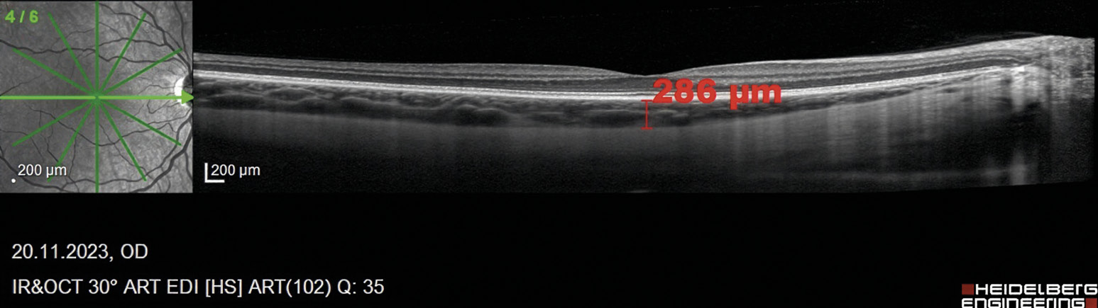
The primary outcome was the subfoveal choroidal thickness (SfChT), measured in micrometers using spectral-domain optical coherence tomography (SD-OCT, Heidelberg Engineering GmbH, Heidelberg, Germany, axial resolution: 3.9 µm/px, repeatability: 7.08 to 34 µm).35–37 The subfoveal choroidal thickness was imaged once by centering a six-line 30° radial scan on the fovea, which was identified at the thinnest point of the foveal area. Extended depth imaging (EDI) function was enabled and the automatic real-time tracking (ART) was set to 100 frames on the SD-OCT to achieve the best possible imaging quality of the choroid. Subfoveal choroidal thickness was determined manually using the distance measurement tool integrated in the device software (Heidelberg Eye Explorer, HEYEX). The distance measurement was performed three times in the 1 : 1 μm view of the horizontal scan line at the same location under the fovea from the end of the Bruch‘s membrane to the end of the choroidal stroma (Figure 2) and the values were averaged. A follow-up was set up in the device software to allow measurements to be taken again at the same retinal location.
To ensure that the data were analyzed as unbiased as possible, the measurements were performed at the end of the study by two blinded examiners. The examiners were trained in the use of the devices and in performing such measurements.
Secondary Outcomes
Axial length and non-cycloplegic objective refraction was measured with Oculus Myopia Master (Oculus Optikgeräte GmbH, Wetzlar, Germany, AL repeatability: 0.02 to 0.14 mm).38–40 Axial length was automatically obtained as an averaged value from a series of six individual measurements weighted according to the signal-to-noise ratio (SNR). It was ensured that the SNR was a minimum of 10 for a correct measurement.
Experimental Setup and Light Exposure
After the initial assessment of the outcome measures, the participants were positioned in front of the lightbox at the correct distance from the light source (Figure 3). The illumination was performed without correction (spectacles or contact lenses), as the examination did not include any fixation or reading tasks and possible spectral distortions caused by lenses could be ruled out. Additionally, it was previously shown that there were no significant differences in axial length following LED red light exposure, regardless of whether the participants were corrected or not.41 After completely darkening the room, participants were instructed to look straight into the light box with red or white light for 20 minutes, depending on the group assignment. 20 minutes were chosen as other studies used comparable durations and it is considered short-term exposure.42 Participants were instructed to look at the diffuse light and not to close their eyes during illumination except to blink. After 20 minutes of illumination, measurements of objective refraction, axial length and subfoveal choroidal thickness were immediately repeated in the same order. To avoid the effects of room lighting, the room remained darkened during the entire study visit. All measurements were taken within two minutes after light exposure.
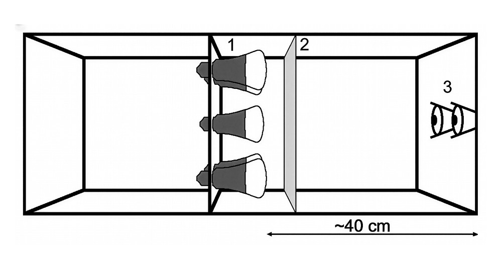
The light exposure was performed in a lightbox that provided direct illumination. The box had a depth of 1 m and a viewing size of 37 × 40 cm (width × height). Five Philips Hue smart bulbs (philips-hue.com) were located on a plane in the box, which was covered by a diffusion foil (LEE Filters 216 White Diffusion, leefilters.com) to create diffuse light. The distance between the LEDs and the corneal surface of the participant was approximately 40 cm (Figure 4).
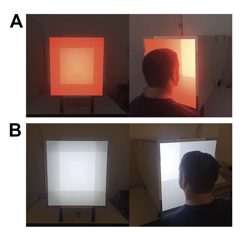
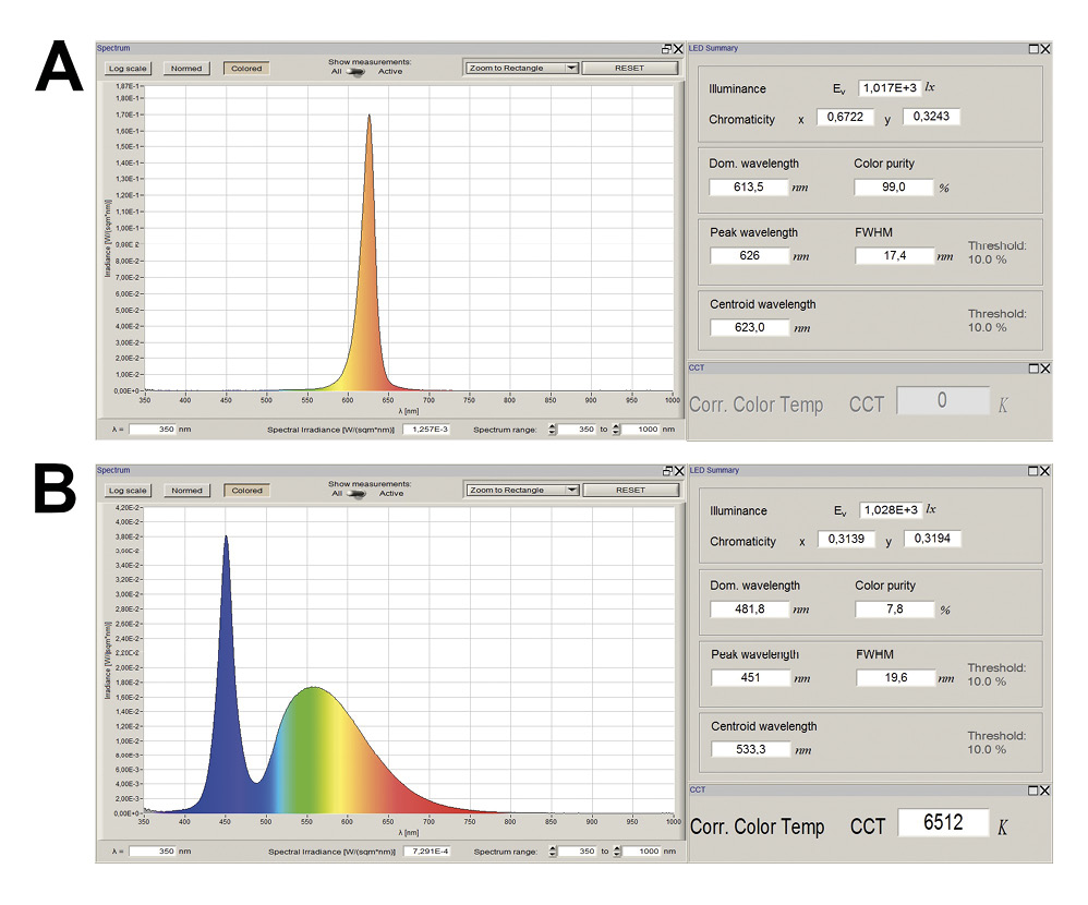
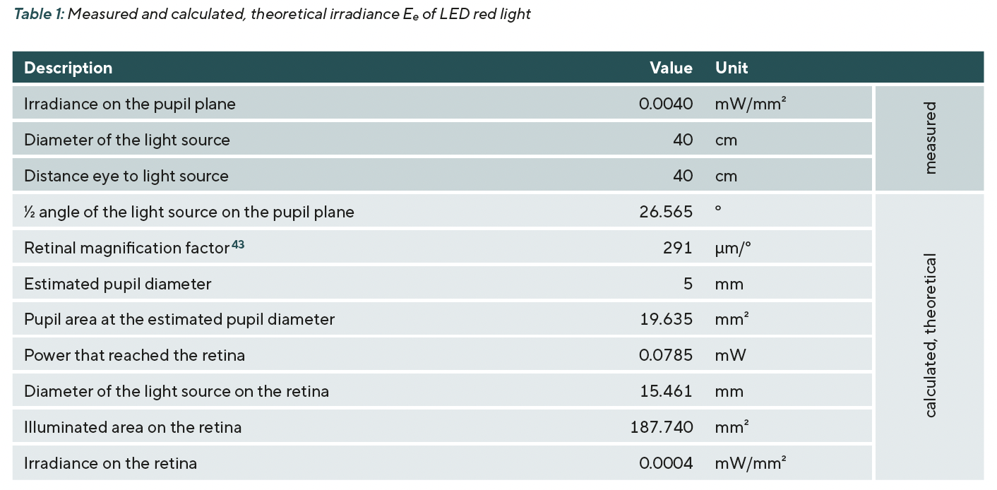
The red light emitted by the LEDs had a wavelength of 623 ± 17 nm (Figure 5A). White light with a color temperature of 6500 K was selected, as this corresponds to the daylight color temperature according to the CIE. Figure 5B illustrates a polychromatic spectrum for white light with a peak at blue wavelengths. This occurs because the desired color temperature cannot be adjusted differently with LEDs based on a fluorescent dye. For both stimuli, an illuminance Ev of 1000 lx was set vertically at eye level at the viewing distance of 40 cm. This corresponds to an irradiance Ee of 0.0040 mW/mm² for red light and 0.0035 mW/mm² for white light at the pupil plane (for the measured and calculated theoretical irradiances of the red experimental illumination, see Table 1). The spectral composition of the light and its intensity were measured using the spectroradiometer JETI specbos 1211 (jeti.com). The selected illuminance corresponds to the minimum requirements for certain activities at the workplace according to the technical rules for workplaces (Technische Regeln für Arbeitsstätten, ASR A3.4) of the Committee for Workplaces (Ausschuss für Arbeitsstätten, ASTA) of the Federal Institute for Occupational Safety and Health (Bundesanstalt für Arbeitsschutz und Arbeitsmedizin, BAuA) Germany.
Statistical Analysis
Statistical analysis was performed using IBM SPSS Statistics Version 29 (IBM Corp. Released 2022. IBM SPSS Statistics for Windows, Version 29.0. Armonk, NY: IBM Corp). Test decisions were made at a significance level of α = 0.05. The outlier analysis was performed visually using boxplots. The Shapiro-Wilk test was used to check for normal distribution, which showed normal distribution for all parameters (p > 0.05). A paired-samples t-test was used to examine the differences in the parameters before and after the respective exposure modality. In addition, the paired-samples t-test was used to examine the differences in these changes between red and white light. Data are presented as mean ± standard deviation unless otherwise mentioned. A one-way ANOVA was performed to examine whether changes differed across refractive groups.
G*Power 3.1.9.7 was used for sample size calculation.44 Based on the results of the study by Swiatczak and Schaeffel (axial length change −13.3 ± 17.3 µm), a minimum required sample size of n = 24 was determined with a power of 95 % and an alpha error probability of 0.05.41 With an estimated drop-out of 20 %, a minimum sample size of n = 30 participants was defined.
Results
Baseline Parameters
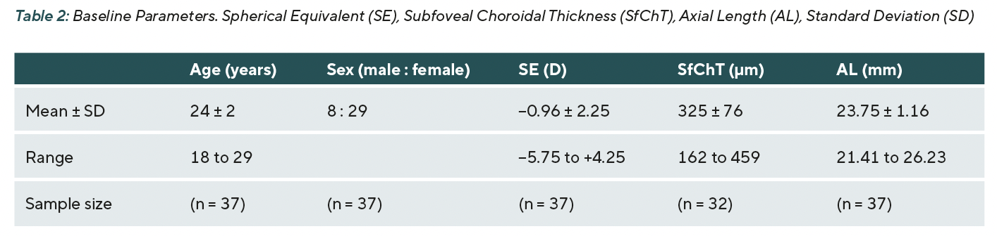
The sample consisted of 37 Caucasian participants (8 male, 29 female) aged 18 to 29 years (mean age 24 ± 2 years) with a mean SE of −0.96 ± 2.25 D (range −5.75 to +4.25 D). Of the 37 participants 22 were individuals with myopia (SE ≤ −0.50 D), 8 were individuals with hyperopia (SE ≥ +0.50 D) and 7 were individuals with emmetropia (+0.25 ≥ SE ≥ −0.25 D). Baseline parameters were derived from the measurements before light exposure of the first appointment of each participant (Table 2).
SfChT before red light exposure was 323 ± 78 µm and before white light exposure 323 ± 77 µm showing no statistically significant differences (mean difference −1 ± 11 µm, p = 0.801). Similarly, AL showed no statistically significant differences (mean difference 0 ± 0.02 mm, p = 0.122) with values before red light exposure of 23.75 ± 1.16 mm and before white light exposure of 23.76 ± 1.17 mm. Overall, this indicates that baseline values for both SfChT and AL did not differ prior to the appointments.
Reliability of Subfoveal Choroidal Thickness Measurement
Intra-rater reliability was evaluated using intraclass correlation coefficient (ICC). The measurements for subfoveal choroidal thickness demonstrated very good reliability with an ICC of 0.997 for rater one and 0.995 for rater two, indicating consistent measurements within each rater.
Inter-rater reliability was evaluated using a Bland-Altman plot.45 The mean difference between raters was 1.34 ± 1.27 µm across all cases. The limits of agreement ranged from +3.83 µm (upper limit) to -1.15 µm (lower limit), suggesting agreement between both raters.
Subfoveal Choroidal Thickness
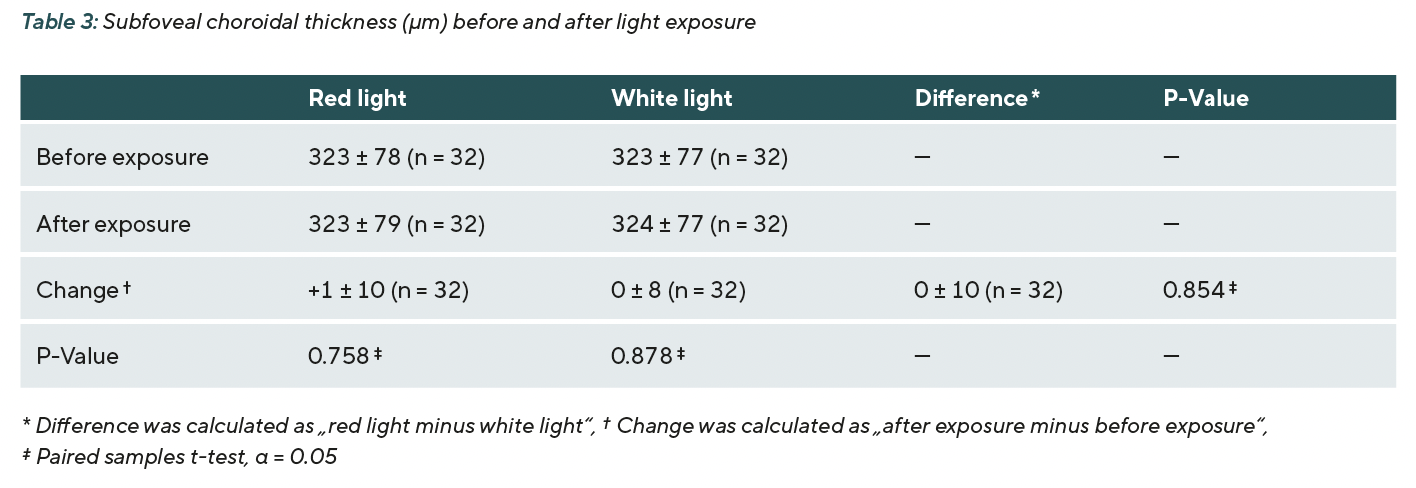
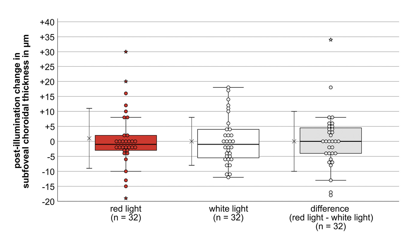
Of the 37 participants, five had to be excluded for choroidal thickness because the image quality was too poor for analysis. Before exposure, the mean SfChT was 323 ± 78 µm and 323 ± 77 μm for red and white light, respectively. After exposure, SfChT did not change for red and white light (mean SfChT: 323 ± 79 μm and 324 ± 77 μm, respectively). Changes due to exposure to red and white light were not statistically significant (mean change: +1 ± 10 μm, p = 0.758 and 0 ± 8 μm, p = 0.878, respectively). The difference between the changes in the experimental and control illumination was also not statistically significant (0 ± 10 μm, p = 0.854) (Table 3). Changes in SfChT showed large individual variability after both red and white light exposure. However, outliers were observed only under red light (Figure 6). No systematic relationship could be found with regard to the outliers or the large variability.
Axial Length
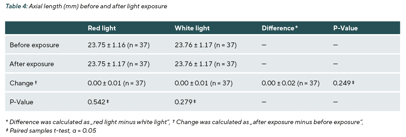
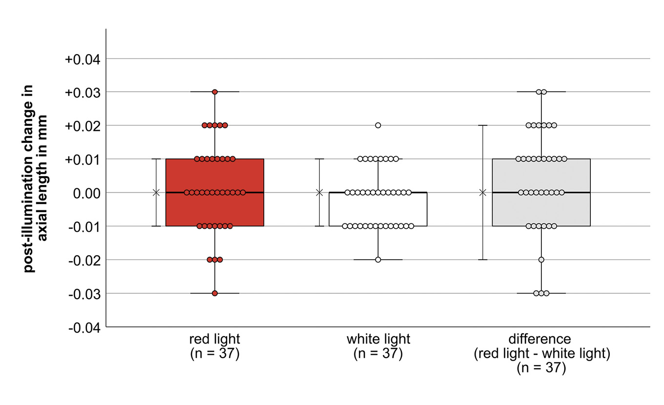
The mean axial length before exposure to red light was 23.75 ± 1.16 mm and before white light 23.76 ± 1.17 mm. After red and white light exposure, the mean axial length did not change (mean AL: 23.75 ± 1.17 mm and 23.76 ± 1.17 mm, respectively). The changes were not statistically significant (mean change after red light: 0.00 ± 0.01 mm, p = 0.542; after white light: 0.00 ± 0.01 mm, p = 0.279). The difference of the changes was also not statistically significant (mean difference: 0.00 ± 0.02 mm, p = 0.249) (Table 4, Figure 7).
Effect of Refractive Error on Subfoveal Choroidal Thickness and Axial Length
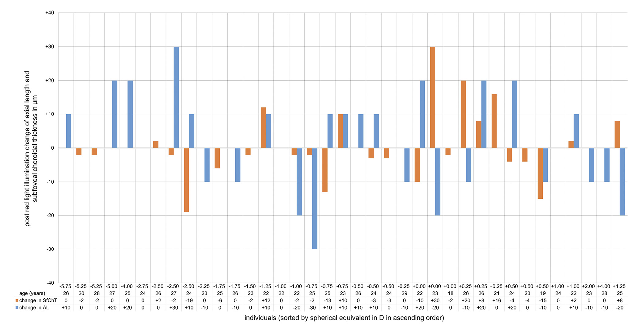
Spherical Equivalent did not statistically significant change after red light (mean change: +0.01 ± 0.29 D, p = 0.886) or after white light (mean change: −0.03 ± 0.24 D, p = 0.405). The difference of the changes was not statistically significant (mean difference: −0.04 ± 0.41 D, p = 0.552). Participants were divided into three groups of ametropia depending on baseline SE: individuals with myopia (n = 22, SE ≤ −0.50 D), individuals with emmetropia (n = 7, +0.25 ≥ SE ≥ −0.25 D) and individuals with hyperopia (n = 8, SE ≥ +0.50 D). One-way ANOVA revealed no significant differences in subfoveal choroidal thickness change between the three refractive groups under either LED red light (p = 0.234) or LED white light (p = 0.487). Similarly, there were no significant group differences in axial length change under LED red light (p = 0.576) or LED white light (p = 0.215). Figure 8 shows the individual responses of participants to subfoveal choroidal thickness and axial length after red light exposure.
Adverse Events
There were no adverse events. No changes or abnormalities were found in the OCT after light exposure, and afterimages caused by the lighting disappeared after the examination.
Discussion
This study investigated the short-term effects of direct LED red light exposure on subfoveal choroidal thickness and axial length. The results show that 20 minutes of direct illumination with LED red light with a wavelength of 623 ± 17 nm and an irradiance of 0.0040 mW/mm² (illuminance of 1000 lx) in a lightbox did not lead to statistically significant changes in the parameters mentioned.
Wavelength and power of red light
Studies on red light laser therapy have shown that its long-term application causes the choroid to thicken and the axial length to decrease.24–26 One publication reported an illuminance of 1600 lx.46 This illuminance is higher than the illuminance used in the current study with LED red light (1600 lx vs. 1000 lx). However, this difference is unlikely to explain the deviating results because for a laser, the power is the relevant factor, which is up to 2 ± 0.5 mW for the RLRL/LLRL systems.22 Such power, which is assigned to laser class 3R, presumably leads with regular use to damaging or inflammatory processes, that could cause the choroid to thicken. Therefore, the wavelength alone does not appear to be a decisive factor for the therapeutic effect in the context of myopia management.
Ambient lighting versus direct illumination
Lou and Ostrin had previously come to different conclusions: After one hour of exposure to ambient LED red light (wavelength: 630 ± 20 nm) there was a significant thinning of the choroid (−5.91 ± 5.72 μm) and a significant increase in axial length.30 The study by Thakur et al. also showed similar results: After one hour of exposure to ambient LED red light (wavelength: 620 ± 35 nm), the choroid thinned significantly (before exposure: 157 ± standard error 8 μm versus after exposure: 146 ± standard error 7 μm) and the eye length increased significantly (+6.4 ± standard error 2.4 μm).31
In both studies, the effect on choroidal thickness was small. It should be noted, particularly in the case of Thakur et al., that choroidal thickness was not measured using OCT but rather using a biometric method that differs from an OCT-based method by 4 ± 24 µm and thus probably does not provide sufficient accuracy to detect the minor effects of red LED light.47 The method for determining choroidal thickness used by Lou and Ostrin is comparable to that used in this study. A main difference between the two cited studies and the current study was the study design. Both studies investigated a longer exposure duration compared to the current study (1 hour versus 20 minutes). Furthermore, the cited studies carried out light exposure with ambient lighting compared to direct light. In the case of Lou and Ostrin‘s study, this resulted in an irradiance 3.5 to 4 times lower than in this study (1 W/m² compared to 4 W/m² for red light and 1 W/m² compared to 3.5 W/m² for white light). This suggests that the exposure duration is a reasonable explanation for the different results, as an effect was found despite lower irradiance. However, the time of day at which the exposure took place, in conjunction with the exposure duration, is a more likely reason for the changes found in red light. Axial length and choroidal thickness underlie physiological and approximately antiphasic fluctuations throughout the day. While axial length increases in the morning, choroidal thickness decreases.33,34 With an exposure between 8:00 a.m. and 11:00 a.m., as used in these studies, the diurnal variation in choroidal thickness could interfere with the effect of light exposure on choroidal thickness and axial length, thus explaining the effect of red light.
The additional findings of the cited studies, that less thinning or even thickening of the choroid and less increase or even decrease in axial length was observed with blue light compared to red light, could presumably be related to the effect of blue light on the (intrinsically) photosensitive retinal ganglion cells (ipRGCs). These cells have a main sensitivity in the range of 480 nm 48 and the blue light of the studies had wavelengths of 456 ± 25 nm and 455 ± 25 nm, respectively. This could have affected the aforementioned diurnal rhythm of axial length and choroidal thickness.49
Irradiance or illuminated retinal area
Swiatczak and Schaeffel recently showed in a similar study that the axial length of 13 non-myopes compared to 17 myopes decreased significantly (−13.3 ± 17.3 µm versus +0.2 ± 12.1 µm, p = 0.02) after 10 minutes of LED red light (wavelength of the filter 620 ± 10 nm, irradiance of 0.0001 mW/mm² at the pupil plane and 0.0025 mW/mm² at the retina, assuming a pupil size of 5 mm and a pupil area of 19.6 mm²).41 Although this irradiance at the pupil plane is 40 times lower than that in the current study (0.0001 mW/mm² versus 0.0040 mW/mm²), theoretically the intensity per mm² on the illuminated retinal area is significantly higher (0.0025 mW/mm² for an area of 0.776 mm² versus 0.0004 mW/mm² for an area of 187.740 mm²). This difference on the retina is explained by the fact that in the current study not only a small spot, but a larger area was illuminated due to the larger light source (3 cm diameter versus 40 cm diameter). Another recent study by Jayakumar with an irradiance at the pupil plane twice as high as in this study (and thus 80 times higher than the study by Swiatczak and Schaeffel) also found no significant differences in axial length after 5 minutes of exposure to LED red light in the morning as well as in the evening with a device (Colordome, Espion, Diagonosys LLC, diagnosysllc.com) that also appears to illuminate a larger retinal area (Jayakumar JPJ, et al. IOVS 2024;65:ARVO E-Abstract 6622). The different results could be caused either by the higher irradiance per area or by the smaller area on the retina that was illuminated, or by a combination of both variables. While significant changes in axial due to red light were found in non-myopic but not in myopic individuals in Swiatczak and Schaeffel’s study, no such change was found in our study. The aforementioned different light modalities (irradiance, stimulus size) could be a reason for this.
Difficulties in investigating short-term effects
These contradictions indicate that there is still no clear explanation of how light affects the regulation of eye growth in humans and which factors, such as intensity, contribute to what extent. The mean deviations of the investigated parameters found in this study are very close to or outside the measurement accuracy of the devices used (SPECTRALIS: 7.08 to 34 μm 35–37, Myopia Master: 0.02 to 0.14 mm 38–40), which means that in most cases, it was not possible to distinguish between measurement error of the device and an effect that may have occurred due to light exposure. In addition, the acquisition of choroidal thickness data from SD-OCT images by manual measurement may be subject to measurement errors. Although Twa et al. have shown that the differences between manual and automatic measurement methods of choroidal thickness in the central retina are not significant, the results of the manual measurement method could vary more between different examiners, especially in cases with thicker choroid where the choroidal-scleral interface is more difficult to detect.50
Limitations
The results of this study and their interpretation must be considered in the context of several limitations:
This study was not conducted in children with growing eyes and it is unclear if these findings in adults with stable refractive errors are applicable. Furthermore, this study examined only visible long-wavelength red light and white light. No conclusions can be drawn regarding the effect of direct light with shorter or longer wavelengths in the visible or non-visible spectrum. Potential influencing factors, such as physical exertion or stress, were assessed via self-report but not objectively controlled. Although the lightbox was homogeneously illuminated and no fixable object was presented, accommodation could not be completely ruled out, which could have influenced choroidal thickness and axial length. However, this applies for both red and white light, which means that the potential influence would probably negligible. In addition, as no fixable object was provided, form deprivation may have affected both parameters. High luminance levels inhibit the development of form deprivation myopia. However, it remains unclear whether this also applies at an illuminance of 1000 lx.51 Furthermore, a hyperopic/myopic defocus could also have influenced the results due to the lack of correction during illumination. Although the measurements were performed immediately after light exposure (with a maximum delay of no more than two minutes), it is still possible that the potential effects of the light stimulus were reduced or no longer visible at the time of measurement. Another limitation could be the measurement of choroidal thickness. This was measured manually by two trained raters. In preliminary studies, several automated methods for measuring choroidal thickness were tested. However, at the time the study was conducted, the automated methods tested were not sufficiently precise in terms of segmentation and repeatability.
Conclusions
The direct LED light used with the investigated exposure modalities showed neither a statistically significant nor a clinically relevant short-term effect on subfoveal choroidal thickness and axial length. Contradictory results from previous studies show that the effect of (red) light in the context of spectral composition, chromaticity, intensity, size of the illuminated area and exposure duration requires further research. However, it remains unclear whether, and under what conditions, exposure to red LED light can have a clinical effect on axial length and choroidal thickness, and thus may have a therapeutic effect in the context of myopia control.
Conflict of Interests
J. Dolata and P. Hessler are lecturers at the academy of Oculus Optikgeräte GmbH. K.-F. Bochenek has no conflict of interests regarding the methods and devices mentioned in the article.
Schroedl, F., Kaser-Eichberger, A., Zhou, X., Wildsoet, C. F. (2023). IMI-The Dynamic Choroid: New Insights, Challenges, and Potential Significance for Human Myopia. Invest. Ophthalmol. Vis.l Sci., 64, 6, 4.